


Our experienced eye doctors, ophthalmic technicians and assistants are ready to provide you with the most comprehensive eye examination possible. Each eye examination will be customized depending on the age group, medical/visual history, and issues of concern for the patient.
The Spectrum Eye Care incorporates visionary digital diagnostic equipment into all of its eye care services. We believe this diagnostic technology is an integral part of our standard of care and helps our doctors and technicians make the right medical decisions for our patients.
Listed below are the medical tests performed in most comprehensive eye examinations.
The Exam A brief overview
1. Hi-technology Pre-Testing
- auto-refraction (refractive error estimate)
- auto-keratometry (cornea curvature)
- non-contact tonometry (glaucoma)
- pachymetry (cornea thickness with Pentacam digital retina scan 21.8 meg Canon and Optomap
2. Medical and Visual Case History
- complete personal and family medical/visual history
3. Visual Acuities (VA)
- distance and near VA - corrected and uncorrected
4. Subjective Refraction
- glasses prescription for distance and near
5. Binocular Vision Testing
- cover test
- near point of convergence
- broad H test
- stereoacuity
6. Colour Vision Testing
- usually performed on pediatric patients or any patient when medically necessary
7. Slit Lamp Biomicroscopy
- microscopic examination of anterior ocular structures - cornea, lens, conjunctiva, etc
8. Direct Ophthalmoscopy
- internal examination of ocular structures, i.e. retina, optic nerve, vitreous, etc.
9. Dilated Fundus Exam with Binocular Indirect Ophthalmoscopy
- internal examination of ocular structures, i.e. retina, optic nerve, vitreous, etc.
- patient pupils are fully dilated (using drops) to allow greater viewing area. performed when medically necessary for at-risk patients
10. Automated Visual Field Testing
- automated test to measure central and peripheral vision performed when medically necessary for at-risk patients
11. Patient Counselling
- discuss examination results with and provide a strategic plan of action with a medical recommendation
12. Patient transfer to Dispensary or Contact Lens Assistants
- eyeglasses and contact lens purchase
AN EXAMPLE OF THE TECHNOLOGY AT THE SPECTRUM
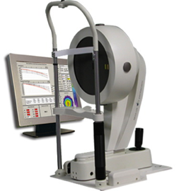
PENTACAM HR The gold standard in analyzing the cornea.
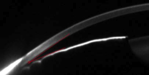
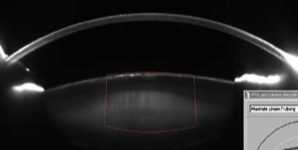
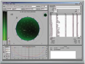
Overview The Pentacam is the gold standard for diagnosis in the anterior eye segment. It supplies topographic data on elevation and curvature of the cornea. The anterior and posterior surfaces are measured from limbus to limbus. The corneal thickness (pachymetry) is also represented graphically over its entire surface.
Very easily we determine the thinnest point of the cornea and all other irregularities quickly and reliably. Using the data on the anterior corneal surface, keratoconus detection is performed.
Important parameters such as anterior chamber angle, depth and volume are calculated and represented automatically by the software. After manual entry of the tonometrically measured intraocular pressure (IOP), the corrected IOP value is calculated and displayed taking the corneal thickness into account.
Gaucoma screening
The Pentacam provides a comprehensive and completely automatic analysis of the anterior chamber. Immediately after the eye has been examined, the instrument displays whether the patient has an increased risk for glaucoma. Post-operative evaluation of the anterior chamber shows alterations, e.g. after an iridectomy or other surgical interventions.
Cataract
The Scheimpflug images produced by the Pentacam supply a clear representation of lens opacity. The 3D cataract analysis combined with the PNS (Pentacam Nucleus Staging) is a unique feature. The centre of the cornea and its anterior and posterior surfaces are measured very precisely for optimal calculation of the refractive corneal power. For your patients this means a perfect calculation of the IOL power – even after refractive surgery.
Contact Lens Fitting
Applications:
Automatic display of all necessary measurement data for fitting contact lenses
Automatic suggestions for contact lenses
Realistic fluo image simulation
Integrated, expandable data bank with over
65,000 lens geometries
With dynamic fluo image simulation, the fit of the contact lens from the intergrated database can be viewed. The simulation makes it possible to adjust inclination and to shift the contact lens while automatically making a new fluo image calculation. The integrated and expandable data bank contains over 65,000 lens geometries. The contact lens geometries can be adjusted individually in cases where fitting is difficult. The user can establish his own rating list for contact lens manufacturers and can expand the database with new or further contact lenses.
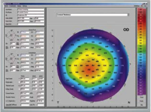
Pachymetric and topographic maps
Pachymetric and topographic maps for individual health care measures Comprehensive refractive maps for transparent diagnostics
Belin/Ambrosio Enhanced Ectasia Display
Extremely sensitive early keratoconus detection and evaluation
Reliable detection of a forme fruste keratoconus in very early stages