By employing different digital filters, we are enabled to detailed views of the RNFL and ILM, highlighting retina folds, cysts, and epiretinal membranes.
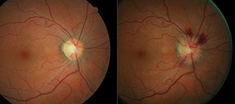
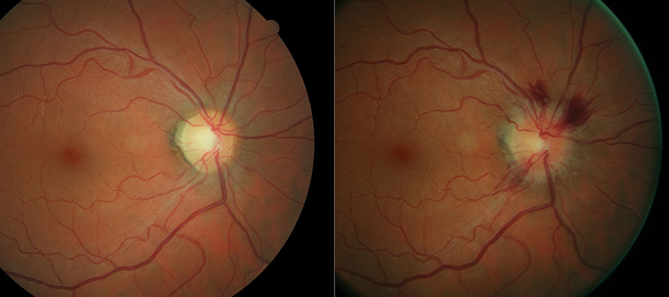
In addition we can focus on the retinal vasculature and highlights common findings, such as hemorrhages, drusen, and exudates. We can also see the choroid, which is useful for visualizing pigmentary disturbances, choroidal ruptures, choroidal nevi, and choroidal melanomas.
In summary we see the vascular structure of the retina as it relates to glaucoma, diabetic retinopathy, and hypertension and by using Fundus autoFluorescence document changes in the Retinal Pigment epithelium (RPE), which may be an early indicator of vision problems, including AMD, glaucoma, diabetic retinopathy, and geographic atrophy.
.
By monitoring subtle Lipofuscin build up in the retina, we are able to assess and monitor the condition of the Retinal Pigment Epithelium (RPE) especially effective in identifying and monitoring subtle changes, even before there’s structural change or vision is affected.
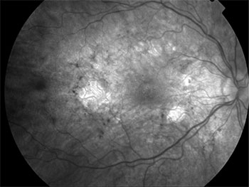
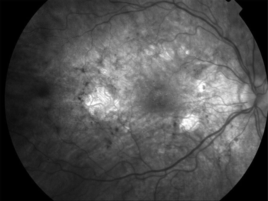
Widespread perimacular RPE atrophy are distributed in this left eye. In the fovea drusen like changes are present.
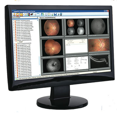
A complete history is kept of all your digital images and they are available to the Drs throughout the two offices locations aiding in a an accurate diagnosis.
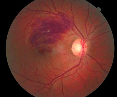
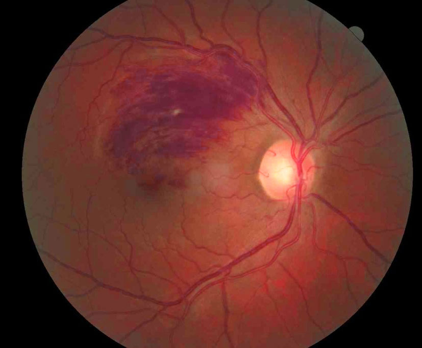
Branch Retinal Vein Occlusion as seen in a complication with diabetes